Physicist Embraces Phages
Fall
2015
In the News
Physicist Embraces Phages
Sigma Pi Sigma alumna and her students dig for new tools to treat disease
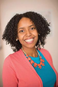
By:Krystle J. McLaughlin, Sigma Pi Sigma Colgate University Chapter, Class of 2004 Professor of Practice, Biological Sciences, Lehigh University
 At the turn of the 20th century, doctors used viruses called bacteriophages to treat bacterial infections. No one knew exactly how the phage therapy worked, and interest in the field stagnated after the advent of antibiotics, which were more effective.
Scientists have taken a renewed interest in phage therapy, as we’ve seen a rise in antibiotic-resistant bacteria over the past several decades. For example, previously treatable bacterial diseases such as tuberculosis can no longer be cured with antibiotics. We are trying to understand the molecular intricacies of how phage infect bacteria and how we can use this knowledge to effectively treat life-threatening bacterial diseases.
Bacteriophages are found in high concentrations wherever their bacterial hosts live, such as in the soil, animal intestines, or the ocean. In just one milliliter of ocean water there are almost a billion virus particles. After infecting their bacterial hosts, the bacteriophage uses the bacteria’s own biological machinery to make copies of itself, filling the bacteria with more phage viruses. The end of this infection culminates in the lysis (a.k.a. explosion) of the bacteria and the release of lots of new bacteriophages that will go on to infect and kill other bacterial cells.
At Lehigh University, in partnership with the Howard Hughes Medical Institute’s Science Education Alliance, Phage Hunters Advancing Genomics and Evolutionary Science (HHMI SEA PHAGES) program, we offer a research-focused course in which undergraduates collect bacteriophages. Each student first brings in a phage-laden soil sample from a location of their choosing. Over the course of the semester, phages that infect a specific bacterial strain are isolated from the soil. Each student names the bacteriophage they’ve isolated (leading to interesting bacteriophage names such as PopTart, Khaleesi, Megatron, and LeBron). Several of the newly discovered phages are characterized by sequencing. The students annotate and analyze phage genomes as part of their course.
In collaboration with the Ware Lab, the undergraduate researchers in my lab group work on one of the phages isolated in the Lehigh SEA PHAGES program. It infects a “cousin” of tuberculosis called Mycobacterium smegmatis, producing several proteins that are unique and do not resemble any other known proteins. Those novel proteins, called orphams, are the targets of our structural characterization. What do they look like? What do they do? We can get insight into their function by resolving the structure with crystallography, as a protein’s structure is intimately related to its function. Students learn how to purify these individual orpham proteins using multiple chromatography methods and then investigate the optimal conditions that will lead to crystallization. Excitingly, these orphams could hold the key to understanding bacteriophage infection and specificity, given their uniqueness.
Our goal is to expand our phage toolbox to allow us to impact treatment of drug-resistant bacterial infections. Perhaps one day we will be able to engineer bacteriophages that will kill bacteria more effectively!
My own interest in biophysics started with the research experiences I had while still a physics undergraduate at Colgate University. I worked on a terahertz spectrometer (I liked that), used an atomic force microscope to image surfaces (very fun!), and studied how cholesterol moved in artificial cell membranes with fluorescence (awesome!).
I enjoyed this last project the most. With the ink still drying on my bachelor’s degree, I started as a biophysics graduate student at the University of Rochester, unsure of what exactly I wanted to study. Then came macromolecular crystallography. I fell in love.
Crystallography let me see the unseeable—coaxing biological macromolecules (e.g., proteins and DNA) into forming crystals, exposing those crystals to a beam of high-energy x-rays, and then dissecting the resulting diffraction patterns through some fun math (reverse Fourier transforms!). The end outcome: a beautiful molecular model.
I loved it and resolved to make it part of my career. Now, perhaps I can pass on that love to my students and make it a part of theirs.
More Information
For more information on the HHMI SEA PHAGES program, including the participating institutions and a complete list of all the phages isolated and named by students (our current count is 6,378, with over 1,000 completed genomes), visit phagesdb.org.
